At our Hertfordshire practice, under the expert care of Dr Joban Sehmi, consultant cardiologist, we provide comprehensive echocardiography services using state-of-the-art imaging technology and evidence-based diagnostic protocols. Our patient-centred approach ensures accurate heart assessments, timely diagnosis, and personalised treatment recommendations delivered with compassionate, expert care.
What Is Echocardiography?
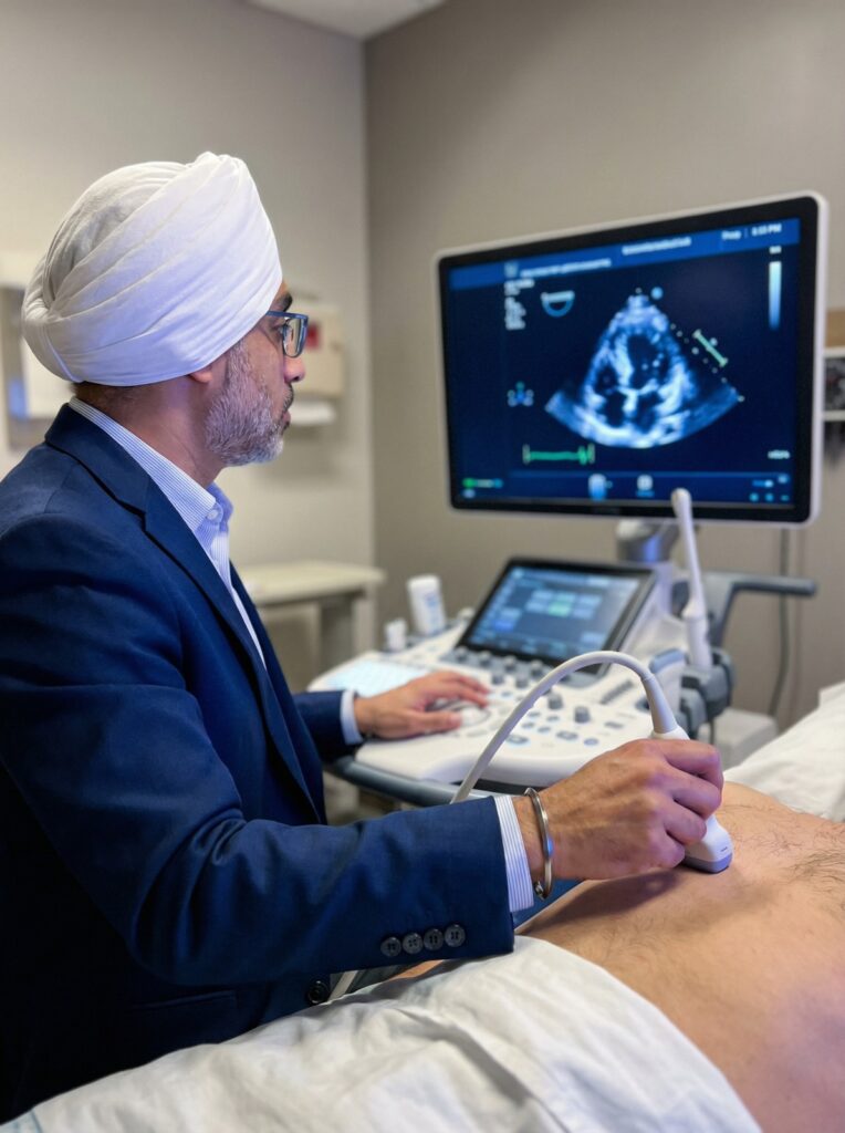
Echocardiography is a sophisticated cardiac imaging technique that uses sound waves (ultrasound) to create detailed moving pictures of your heart’s structure, function, and blood flow. This safe, non-invasive diagnostic tool provides real-time visualisation of your heart’s chambers, valves, and blood vessels without radiation exposure. We offer several specialised types of echocardiography to meet different diagnostic needs, each providing unique insights into your cardiac health.
Types of Echocardiography Services
Transthoracic Echocardiography (TTE)
Transthoracic Echocardiography is the most common type of heart ultrasound, performed by placing an ultrasound transducer on your chest wall. This non-invasive procedure provides comprehensive evaluation of your heart’s structure and function through real-time imaging. The test is painless, requires no preparation, and typically takes 30-45 minutes. TTE serves as the foundation of cardiac assessment and is suitable for patients of all ages, from newborns to elderly adults.
TTE is used to diagnose and monitor:
- Heart valve disease including stenosis and regurgitation
- Heart muscle function and pumping efficiency
- Heart chamber sizes and wall thickness
- Congenital heart abnormalities present from birth
- Pericardial disease including fluid around the heart
- Cardiac masses and tumours
- Pulmonary hypertension and right heart function
- Cardiomyopathy and heart failure assessment
Benefits of Transthoracic Echocardiography:
- Completely non-invasive with no needles or sedation required
- No radiation exposure, safe for repeated examinations
- Immediate real-time imaging and assessment
- Painless and well-tolerated by all patients
- Cost-effective first-line cardiac diagnostic tool
- Suitable for patients of all ages including pregnant women
- Provides comprehensive cardiac structural and functional information
- Convenient outpatient procedure with no recovery time needed

Transesophageal Echocardiography (TOE/TEE)
Transesophageal Echocardiography involves positioning a specialised ultrasound probe in the oesophagus (food pipe) to obtain exceptionally clear images of the heart from within the chest. Because the probe is positioned directly behind the heart without chest wall or lung interference, TOE provides superior resolution and detailed visualisation of cardiac structures that may be difficult to see with standard transthoracic imaging. The procedure is performed under conscious sedation for patient comfort and typically takes 15-30 minutes.
TOE is particularly valuable for diagnosing:
- Atrial fibrillation assessment and stroke risk evaluation
- Detection of blood clots within heart chambers, especially the left atrial appendage
- Heart valve disease requiring detailed assessment before surgery
- Infective endocarditis (heart valve infections) and vegetation detection
- Aortic disease including dissection and aneurysm evaluation
- Congenital heart disease including holes in the heart (septal defects)
- Cardiac tumours and masses requiring precise characterisation
- Prosthetic (artificial) heart valve function assessment
- Guidance for interventional procedures including catheter-based treatments
Benefits of Transesophageal Echocardiography:
- Superior image quality with minimal acoustic interference from chest structures
- Exceptional visualisation of posterior heart structures and left atrium
- Highly accurate for detecting blood clots before cardioversion procedures
- Essential for surgical planning requiring detailed valve anatomy
- Real-time procedural guidance for catheter-based interventions
- Detailed assessment of complex congenital abnormalities
- Precise evaluation of prosthetic valve function and complications
- Enhanced diagnostic confidence for stroke investigation and prevention
- Performed safely under sedation with continuous monitoring
Exercise Stress Echocardiography
Exercise Stress Echocardiography assesses how your heart performs during physical activity, revealing how effectively it functions under natural physiological stress. The test combines standard echocardiographic imaging with treadmill or bicycle exercise to evaluate cardiac reserve and detect coronary artery disease. You will exercise according to a standardised protocol with gradually increasing workload while your heart rate, blood pressure, and ECG are continuously monitored. Images are obtained at rest before exercise and immediately after you reach peak exercise capacity, allowing comparison of heart function during different levels of demand.
Exercise Stress Echocardiography is used to diagnose:
- Coronary artery disease and cardiac ischaemia detection
- Angina investigation and evaluation of exercise-induced chest pain
- Heart attack risk stratification in patients with exertional symptoms
- Assessment of known coronary disease severity and functional impact
- Evaluation of cardiac reserve and true exercise capacity
- Heart valve disease behaviour during physiological exercise conditions
- Exercise-induced arrhythmia assessment and symptoms correlation
- Preoperative cardiac risk assessment in physically active patients
- Functional capacity evaluation for rehabilitation and activity prescription
Benefits of Exercise Stress Echocardiography:
- Physiological stress that mimics real-world activity and symptoms
- Non-invasive alternative to coronary angiography for many patients
- No radiation exposure unlike nuclear stress testing
- Assessment of actual exercise capacity and functional limitations
- High diagnostic accuracy for detecting significant coronary disease during exertion
- Simultaneous evaluation of symptoms, ECG changes, and cardiac function
- Prognostic information for risk stratification and treatment planning
- Safe procedure performed under continuous medical supervision with emergency equipment available
- Helps determine safe exercise levels and guides cardiac rehabilitation programmes
- Immediate results with real-time visualisation during peak stress
Dobutamine Stress Echocardiography
Dobutamine Stress Echocardiography uses medication to simulate the effects of exercise on your heart, providing an alternative for patients unable to exercise adequately due to mobility limitations, arthritis, lung disease, or other physical constraints. Dobutamine is a synthetic medication administered through an intravenous line that increases heart rate and contractility, creating cardiac stress similar to exercise. The medication is given in gradually increasing doses while your heart is continuously monitored with echocardiography, ECG, and blood pressure measurements. Images are obtained at rest and at multiple stages during medication infusion to assess how different areas of heart muscle respond to increasing stress.
Dobutamine Stress Echocardiography is used to diagnose:
- Coronary artery disease in patients unable to exercise adequately
- Cardiac ischaemia detection when exercise testing is not feasible
- Myocardial viability assessment after heart attacks to identify recoverable muscle
- Assessment of coronary disease severity and functional significance
- Evaluation of cardiac reserve in patients with exercise limitations
- Preoperative cardiac risk assessment in patients with mobility restrictions
- Low-dose protocols for viability testing in heart failure patients
- Stress-induced wall motion abnormalities indicating areas of reduced blood supply
Benefits of Dobutamine Stress Echocardiography:
- Excellent alternative for patients unable to perform adequate exercise
- Controlled medication-induced stress with precise dosing titration
- Allows stress testing in patients with orthopaedic, neurological, or respiratory limitations
- Shorter procedure time compared to exercise protocols in some cases
- High diagnostic accuracy comparable to exercise stress testing
- Viability assessment capabilities not available with exercise stress alone
- Safe procedure with immediate medication reversal if needed
- Performed under close medical supervision with continuous monitoring
- No radiation exposure and immediate results available
- Particularly valuable for elderly patients or those with physical disabilities
Contrast Echocardiography
Contrast Echocardiography involves injecting a special contrast agent (microbubbles) through an intravenous line to enhance ultrasound images of the heart. These harmless gas-filled microspheres improve visualisation of heart chamber borders, blood flow patterns, and cardiac structures that may be difficult to see with standard imaging alone. Contrast enhancement is particularly valuable in patients with technically difficult imaging windows or when precise assessment of heart muscle function is essential. The contrast agent is safe, well-tolerated, and cleared rapidly from the body through normal breathing.
Contrast Echocardiography is particularly useful for:
- Enhanced visualisation of heart chamber borders and wall motion
- Improved accuracy in assessing left ventricular function and ejection fraction
- Detection of intracardiac shunts (abnormal connections between heart chambers)
- Myocardial perfusion imaging to assess blood flow to heart muscle
- Evaluation of cardiac masses and structural abnormalities requiring enhanced definition
- Assessment in patients with difficult imaging windows due to body habitus or lung disease
- Stress echocardiography enhancement for improved wall motion analysis
Benefits of Contrast Echocardiography:
- Dramatically improved image quality in technically challenging patients
- Enhanced accuracy in measuring heart function and chamber volumes
- Better visualisation eliminates the need for alternative imaging in many cases
- Safe contrast agent with minimal side effects and rapid clearance
- Improved diagnostic confidence reducing the need for additional testing
- Enhanced detection of subtle wall motion abnormalities during stress testing
- Cost-effective enhancement of standard echocardiography without radiation
- Particularly valuable in obese patients or those with lung disease
Why Choose Dr Joban Sehmi for Ambulatory Blood Pressure Monitoring?
Specialist Consultant Cardiologist Consultant cardiologist with expert training in blood pressure disorders, hypertension management, and cardiovascular risk assessment and treatment optimisation.
Advanced Monitoring Technology Access to the latest digital ambulatory blood pressure monitors providing accurate, validated measurements with comfortable, lightweight design for patient convenience.
Comprehensive Cardiovascular Care Complete diagnostic service from initial assessment through detailed report interpretation, treatment planning, and ongoing blood pressure management.
Expert Report Interpretation Detailed analysis of blood pressure patterns, nocturnal dipping status, and cardiovascular risk assessment with clear, actionable treatment recommendations.
Patient-Centred Approach Compassionate care with clear explanations tailored to your individual concerns, practical guidance on monitor use, and accessible support during monitoring.
Rapid Access Convenient appointment availability with prompt monitor fitting, quick turnaround for results reporting, and timely treatment recommendations.
Evidence-Based Practice Diagnostic and management protocols aligned with NICE guidelines, European Society of Hypertension standards, and international best practice recommendations.
Hertfordshire Convenience Local access to consultant-level blood pressure assessment and hypertension management without the need to travel to distant specialist centres.
Integrated Care Pathway Seamless coordination with other specialists and your GP for comprehensive cardiovascular risk management and treatment optimisation.
Holistic Approach Consideration of blood pressure within your overall cardiovascular risk profile, including lifestyle factors, other risk factors, and existing medical conditions.
Long-Term Partnership Ongoing monitoring and management support for sustained blood pressure control and cardiovascular health throughout your treatment journey.
Clear Communication Plain-language explanations of findings, blood pressure patterns, cardiovascular implications, and recommended next steps you can understand.
Book Your Appointment with Dr Joban Sehmi
Take control of your blood pressure and cardiovascular health today. If you have elevated clinic blood pressure readings, require confirmation of hypertension diagnosis, need assessment of treatment effectiveness, or have concerns about your blood pressure control, expert care is available close to home. Contact Dr Joban Sehmi’s Hertfordshire practice to schedule your ambulatory blood pressure monitoring appointment and benefit from consultant-level diagnostic assessment with personalised, compassionate care.
Your journey to better heart health starts here.